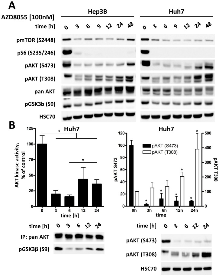
Figure 4.
Inhibition of mTORC1/2 causes upregulation of phospho-AKT at T308, resulting in increased residual AKT activity over time in HCC cell lines. (A) Hep3B and Huh7 cells were treated with either DMSO or 100 nM AZD8055 for up to 48 h and cell lysates were prepared at the indicated time points. PI3K-AKT-mTOR pathway signaling was analyzed by western blot. HSC70 was used as loading control. (B) Huh7 cells were treated with 100 nM AZD8055 for 0, 3, 6, 12 and 24 hours. AKT in vitro kinase assays were performed after quantitative pan AKT immunoprecipitation in triplicates per timepoint. GSK3α/β fusion protein was used as AKT substrate and phosphorylation at S9/21 detected by western blot. pAKT S473 and T308 levels were analyzed by western blot. Bars: SD. *, p < 0.05; #, p < 0.01

