Figure 3.
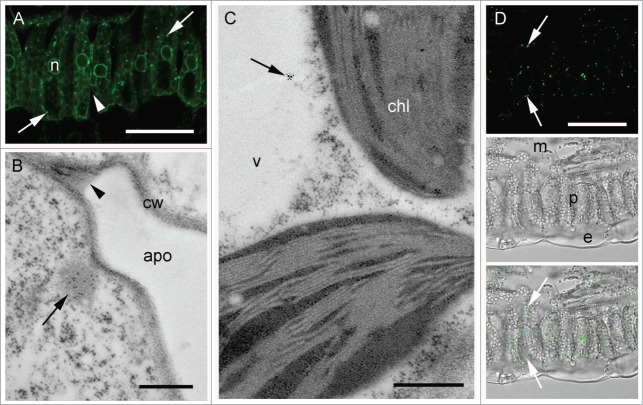
Localization of SIgA complexes in plant leaf sections. (A) Transverse section of palisade mesophyll, after IHC with anti-α chain antiserum and Alexa Fluor® 488-conjugated secondary antibody. Arrows indicate PB-like structures (B) Immunogold labeling with anti-α chain antiserum, of the membrane-proximal region of a mesophyll cell showing apoplast (apo) and cell wall (cw) structures. The labeling is concentrated in the PB-like structures (arrow), whereas no labeling is visible in the apoplast (arrowhead) (C) Immunogold labeling in the vacuole (arrow) with anti-α chain and anti-kappa chain antisera in combination. Vacuole (v) and chloroplast (chl) structures are indicated (D) IHC with an anti-2G12 idiotype antibody and an Alexa Fluor® 488-conjugated secondary antibody. Arrows indicate deposition sites of antibody complexes containing the 2G12 idiotype; Lower panels, phase contrast images of a transverse section of palisade mesophyll with (lower) and without (middle) fluorescence overlay. Scale bars: 100 μm (A), 0.5 μm (B and C), 200 μm (D).

