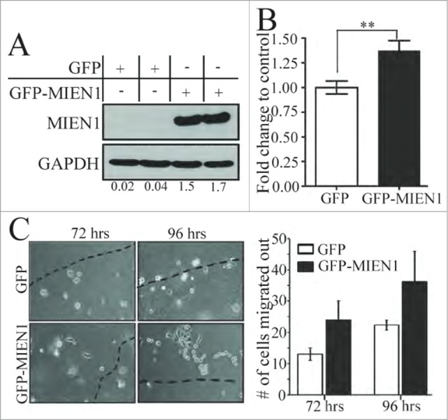
Figure 4.

MIEN1 overexpression in DOK cells results in increased migration and invasion. (A) Western blotting shows MIEN1 expression upon GFP empty vector or GFP-MIEN1 plasmid transfection. (B) Quantification of invasive potential of transfected cells 24 hours after reseeding transfected cells on inserts. (C, left) Representative images of agarose beads at respective time points after reseeding GFP or GFP-MIEN1 transfected cells; (C, right) Quantification of number of cells migrated out from at least 10 independent fields.
