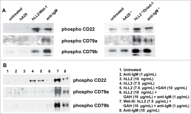
Figure 3.

Phosphorylation of CD79a, CD79b, and CD22. Western blot analyses of phosphorylated CD79a, CD79b, and CD22 in D1–1 cells treated for 2 h with (A) the Wet-I format of soluble antibodies (left panel) or the Dried-I format of immobilized antibodies (right panel), and (B) various formats of soluble epratuzumab, including Wet-I (lane 4; hLL2, 7.5 μg/mL), Wet-IIA (lane 5; hLL2, 7.5 μg/mL; GAH, 10 μg/mL), and Wet-III (lane 7; hLL2, 7.5 μg/mL; GAH, 10 μg/mL; anti-IgM, 1 μg/mL). In lane 6, the amounts of GAH and anti-IgM were the same as those in lane 7, but the concentration of epratuzumab was too low (10 ng/mL) to induce a notable effect.
