Figure 4.

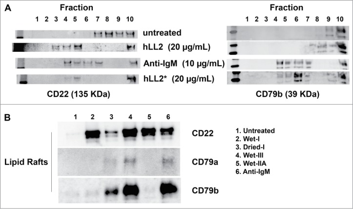
Translocation of CD79a, CD79b, and CD22 to the lipid rafts. (A) left panel, CD22 was detected in the lipid rafts (fractions 3–6) following treatment of D1–1 cells with anti-IgM (10 μg/mL) and both the Wet-I (hLL2, 20 μg/mL) and Dried-I (hLL2*, 20 μg/mL) formats of epratuzumab; right panel, CD79b was translocated to the lipid rafts (fractions 4–7) by either anti-IgM (10 μg/mL) or the Dried-I format of epratuzumab (hLL2*, 20 μg/mL), but not soluble epratuzumab (hLL2). (B) Anti-IgM (lane 6) and epratuzumab of the Dried-I (lane 3) or Wet-III (lane 4) format, but not the Wet-I (lane 2) or Wet-IIA (lane 5) format, induced redistribution of CD22, CD79a, and CD79b to the lipid rafts.
