
Figure 3.
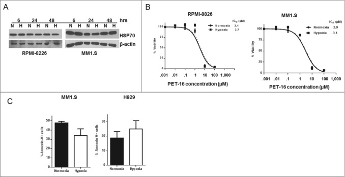
No effect of hypoxia on cytotoxicity by PET-16. (A) MM cells were kept in normoxia (N) or hypoxia (H) for the indicated period of time. Cells were then collected and the level of HSP70 expression was determined by Western blotting. Equal loading was confirmed by re-probing the membrane with antibody against β-actin. The results depicted are representative of multiple independent experiments. (B) MM cells were cultured in the presence of indicated concentrations of PET-16 in normoxia or hypoxia for 72h. Cell viability was measured in triplicate using Resazurin (alamar blue) dye. Error bars mark standard deviations. (C) MM cells were treated with PET-16 (3 μM) for 24 hrs, then collected and apoptosis was determined using Annexin V binding assay. Combined results from 3 independent experiments are shown. Data are presented as mean ± SD.