Figure 1.

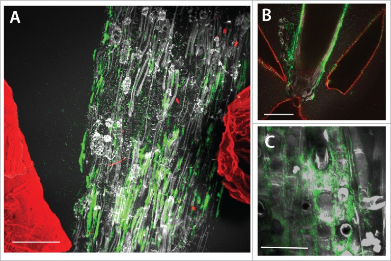
Maximum projection confocal images of GFP-labeled Pseudomonas fluorescens colonies (green) on the surface of lettuce root tissues (gray) in situ in transparent soil with Nafion particles from the substrate labeled with sulphorhodamine B fluorescent dye also visible (red). (A) The majority of the bacterial fluorescence is associated with the root tissue. Scale bar = 150 μm. (B) Bacteria are present on the root tip and in this case also the surfaces of Nafion particles in close proximity to the root have bacterial fluorescence associated with them. Scale bar = 150 μm. (C) At higher resolution, bacterial colonisation was predominantly observed in the intercellular junctions of root epithelial cells. Scale bar = 45 μm.
