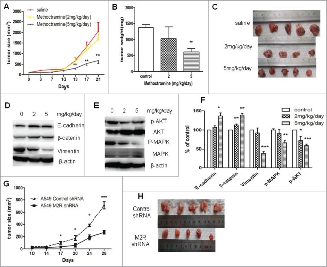
Figure 5.
Effects of blocking M2R signaling on the growth of A549 tumor xenografts in nude mice. (A–C), Methoctramine treatment inhibited tumor growth in a dose-dependent manner. A549 cells were injected s.c. into the left flank of each nude mouse. Methoctramine treatment with indicated dosage started one week after injection. Control group was given the same amount of saline daily. (A) Tumor volume growth curve. Tumor volumes and mice body weight were measured 3 times a week. (B) Tumor weight. Tumors were removed from mice after 3-week treatment and weighed. (C) Photographs of tumors removed from mice after 3-week treatment. (D and E) Western blot showed that 3-week methoctramine treatment decreased MAPK and Akt phosphorylation (D) and reversed EMT (E) in tumor xenografts in a dose-dependent manner. (F) Quantification of Western blots shown in D and E. (G and H) A549 M2 shRNA cells showed a slower growth rate than A549 control shRNA cells in nude mice. Same amount of A549 M2 shRNA cells or A549 control shRNA cells were injected s.c. into the left flank of each nude mice. (G) Tumor volume growth curve. Tumor volumes and mice body weight were measured twice a week. (H) Photographs of tumors removed from mice after 4 weeks. For animal experiment, each group had 4–5 mice and each experiment was repeated twice. Data were shown as mean±s.e.m.*, P < 0.05;**, P < 0.01; ***, P < 0.001, compared with control.

