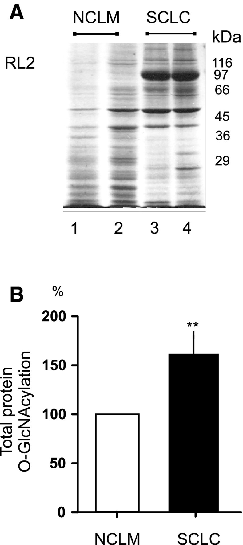
Fig. 3.
Representative image obtained from normal and laryngeal cancer sample homogenates (50 μg protein loaded per lane) probed with the RL2 antibody, where multiple bands were detected and used to quantify protein O-GlcNAcylation (a); plots demonstrate significant increases of ~60 % in total protein O-GlcNAcylation levels in laryngeal cancers tissues compared with normal tissues; **p < 0.01 (b)

