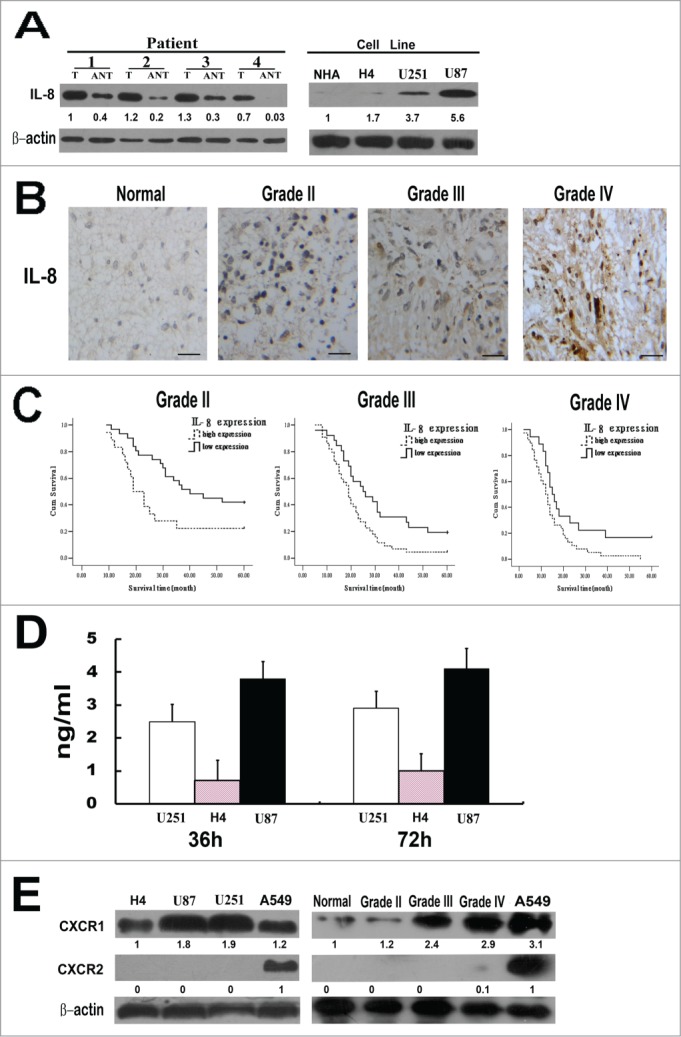
Figure 1.

IL-8 expression in glioma tissues and cell lines. (A) Left, Expression of IL-8 protein in paired glioma tissues (T) and adjacent non-tumor tissues (ANT), with each pair obtained from the same patient. Right, Expression of IL-8 protein in cultured glioma cell lines(normal human astrocytes (NHA) cells, H4, U251 and U87). β-actin was used as a loading control. Quantification of relative protein levels is shown below the blots. The results were from a representative of at least 3 repeated experiments. (B) Expression of IL-8 protein in normal brain tissue and glioma tissues (grade II, grade III and grade IV) was examined by immunohistochemical staining, scale bar: 20 um. (C) The statistical significance of the difference between curves of IL-8 high-expressing (dotted line) and low-expressing (bold line) patients was compared within subgroups of WHO grade II, grade III and grades IV. P values were calculated by the log-rank test, P < 0.05 was considered statistically significant. (D) Glioma cells secrete IL-8 in the complete media. Three different types of glioma cell lines were plated in 96 well plates and culture media was collected at the end of 36 and 72 h for ELISA measurement. The results were from a representative of at least 3 repeated experiments. (E) Expression levels of CXCR1 and CXCR2 protein in glioma cell lines (H4, U87, U251) and in normal brain tissue and glioma tissues (grade II, grade III and gradeIV) were detected by Western blot. A549 cell line was used as a positive control, β-actin was used as a loading control. Quantification of relative protein levels is shown below the blots. The results were from a representative of at least 3 repeated experiments.
