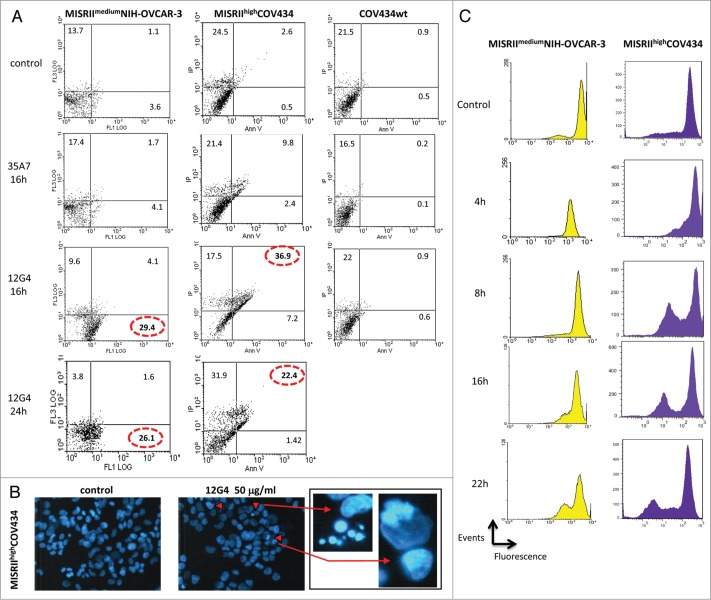
Figure 4.
The mAb 12G4 induces apoptosis of MISRIImediumNIH-OVCAR-3 and MISRIIhighCOV434 cells, but not of COV434 cells. (A) Cells were incubated or not with 50 μg/mL 12G4 for 16 h and 24 h or with 50 μg/mL 35A7 (irrelevant antibody) for 16 h and then the apoptosis rate was quantified by flow cytometry following Annexin V/propidium iodide (PI) staining. (B) Representative fluorescence images of MISRIIhighCOV434 cells incubated or not with 50 μg/ml of 12G4 for 24 h. Apoptotic cells (arrows) have bright blue condensed or fragmented nuclei stained by Hoechst 33258. (C) DIOC6 (fluorescent lipophilic dye) assay at 4 h, 8 h, 16 h, and 22 h after incubation with 50 μg/mL 12G4. The decrease of the fluorescence peak corresponds to the inhibition of the mitochondrial membrane potential following apoptosis.

