
Fig. 8.
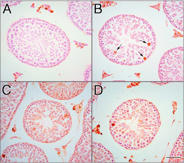
Localization of HSP70 in the EI testes. In the negative control testis, the primary antibody was omitted, and no labeling of germ cells was observed. Nonspecific staining of Leydig cells was observed in interstitial tissues (A). In the control testis, positive labeling was observed in a few spermatocytes (B, arrows). In EI-120 (C) and EI-180 (D) testes, immunoperoxidase staining was also observed in all round and elongated spermatids and spermatocytes. Scale bar, 50 μm.