
Figure 1.
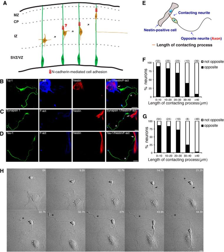
The Nestin-positive cell–cortical neuron interaction directs axon formation from the opposite side of the contacting neurite. A, Schematic diagram of the proposal that RGC–pyramidal cell interaction directs axon–dendrite polarization for the initiation of the subsequent Radial glia-guided neuronal migration. The leading-like process of a multipolar cell may already bind to a radial glial fiber through N-cadherin-mediated cell adhesion, and the binding itself may initiate the MBT. B–D, Representative images of the interaction between Nestin-positive cells (red) and cortical neurons (green) in vitro. Axons form from the side of the cortical neuron opposite the Nestin-positive cell-contacting neurite. Arrows and asterisks indicate the Nestin-positive cell-contacting neurite and nascent axon, respectively. Nestin-positive cells and cortical neurons were cocultured for 2 d, and cells were subsequently immunostained with the indicated antibodies. Tuj-1, neuron marker; Tau-1 and Synapsin-1, axon marker; Nestin, radial glial cell marker; and F-act, stained with Alexa Fluor 647-conjugated phalloidin (blue). Scale bars, 20 μm. E, Schematic diagram of a typical Nestin-positive cell–cortical neuron interaction. Along the somatic polarity axis, the axon forms on the side opposite the Nestin-positive cell-contacting neurite. F, G, Quantitative analysis of the direction of axon formation. The length of the contacting process is indicated in E. The number above each bar indicates the total number of examined Nestin-positive cell-contacting neurons. F, Neurons were labeled with the neuronal marker Tuj-1, and Nestin-positive cells were labeled with F-actin and the radial glia marker Nestin. G, Neurons were labeled with the neuron marker Tuj-1 and the axonal marker Tau-1. Nestin-positive cells were labeled with F-act. H, Time-lapse images of the interaction between cocultured Nestin-positive cells and cortical neurons. Images were acquired every 10 min. Arrows and asterisks indicate the Nestin-positive cell-contacting neurite and the axon, respectively. Scale bars, 10 μm. F-act, F-Actin; MZ, marginal zone; R, radial glial cell; n, pyramidal neuron.