
Figure 2.
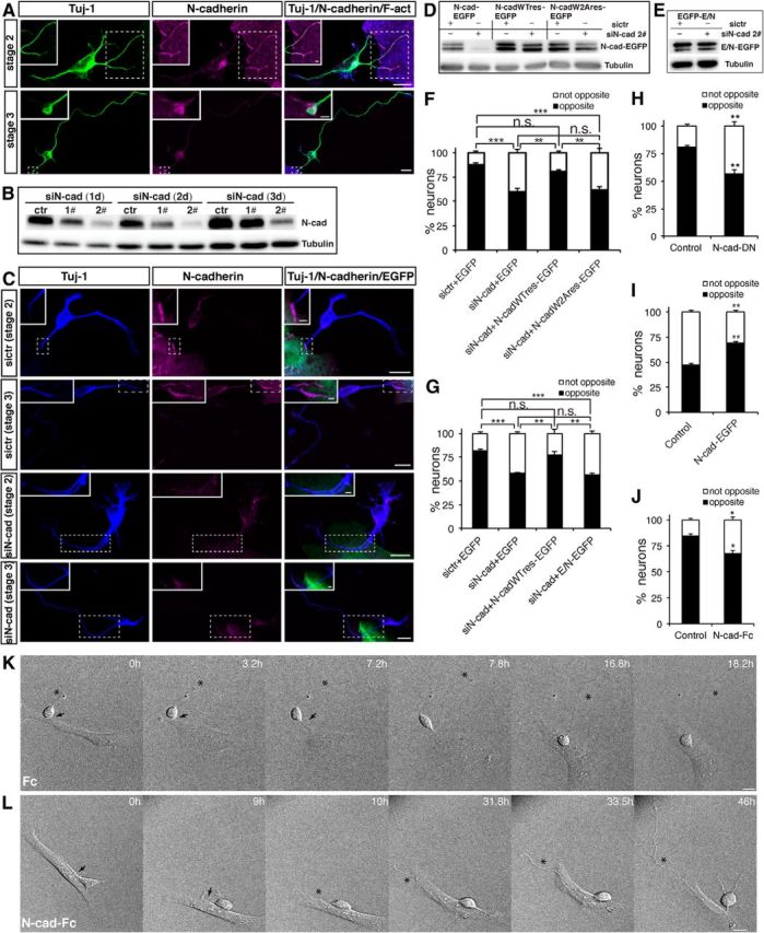
N-cadherin is required for the Nestin-positive cell–cortical neuron interaction in vitro. A, C, The distribution of N-cadherin at the contact sites between neurons and Nestin-positive cells under various experimental conditions. A, Nestin-positive cells and cortical neurons were cocultured for 1 d, and the cells were subsequently immunostained with the indicated antibodies. C, Purified Nestin-positive cells were transfected with control siRNA or siN-cad 2# plus pEGFP. The following day, the transfected Nestin-positive cells were cocultured with dissociated cortical neurons for 1 d, and the cells were subsequently immunostained with the indicated antibodies. Scale bars, 10 μm. Magnification of the boxed region shows the distribution of N-cadherin at the contact site between the Nestin-positive cell and cortical neuron. Scale bars, 2 μm. B, Lysates from purified Nestin-positive cells transfected with the indicated siRNA were subjected to immunoblot analysis using the indicated antibodies. D, E, Lysates from COS-7 cells transfected with the indicated siRNA and plasmids were subjected to immunoblot analysis using the indicated antibodies. F, G, Quantitative analysis of the direction of axon formation. Purified Nestin-positive cells transfected with the indicated siRNA and plasmid were cocultured with dissociated cortical neurons for 2 d, and the cells were subsequently analyzed by immunostaining. Neurons were labeled with Tuj-1, and Nestin-positive cells were labeled with GFP and Nestin. **p < 0.01, ***p < 0.001, Tukey's multiple-comparison test. n.s., Not significant. Error bars represent the SEM. n ≥ 40 Nestin-positive cell-interacting neurons. H, Quantitative analysis of the direction of axon formation. Cortical neurons transfected with pCAG-myc-GST or cotransfected with pCAG-myc-GST and pCAG-MCS2-N-cadherin-DN were cocultured with purified Nestin-positive cells for 2 d, and the cells were subsequently analyzed by immunostaining. Neurons were labeled with Myc, and Nestin-positive cells were labeled with F-act and N-cadherin. **p < 0.01, Student's t test. Error bars represent the SEM. n ≥ 40 Nestin-positive cell-interacting neurons. I, Quantitative analysis of the direction of axon formation. L cells transfected with the indicated plasmid were cocultured with dissociated cortical neurons for 2 d, and the cells were subsequently analyzed by immunostaining. Neurons were labeled with Tuj-1 and Hoechst, and L cells were labeled with GFP and Hoechst. **p < 0.01, Student's t test. Error bars represent the SEM. n ≥ 40 L cell-interacting neurons. J, Quantitative analysis of the direction of axon formation. Nestin-positive cells and cortical neurons were cocultured in the presence of the Fc control or N-cad-Fc for 2 d, and the cells were subsequently analyzed by immunostaining. Neurons were labeled with Tuj-1, and Nestin-positive cells were labeled with F-actin and Nestin. *p < 0.05, Student's t test. Error bars represent the SEM. n ≥ 40 Nestin-positive cell-interacting neurons. K, L, Time-lapse images of the interaction between Nestin-positive cells and cortical neurons under various experimental conditions. Arrows and asterisks indicate the Nestin-positive cell-contacting neurite and the nascent axon, respectively. Scale bars, 10 μm.