
Figure 5.
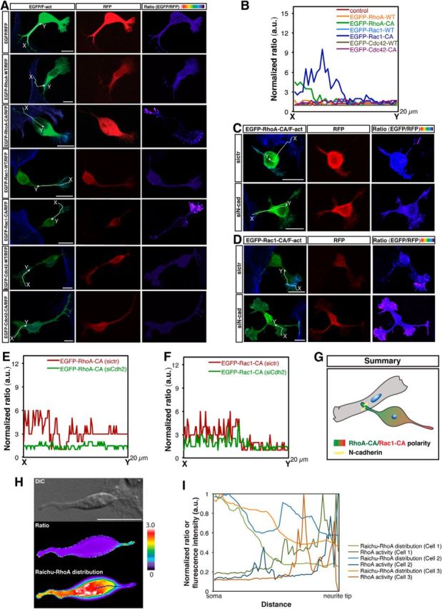
The N-cadherin-dependent Nestin-positive cell–cortical neuron interaction induces polarized distribution of active RhoA at the contact site and active Rac1 in the opposing neurite. A, C, D, Representative images of the spatial distributions of RhoA-CA, Rac1-CA, and Cdc42-CA in cultured Nestin-positive cell-contacting cortical neurons under various experimental conditions. A, Dissociated cortical neurons were cotransfected with the indicated plasmids using in vitro electroporation and cocultured with purified Nestin-positive cells for 1 d. C, D, Purified Nestin-positive cells were transfected with control siRNA or siN-cad 2#. One day later, transfected Nestin-positive cells were cocultured with dissociated cortical neurons cotransfected with the indicated plasmids using in vitro electroporation for 1 d. The fluorescence intensity ratio of EGFP to RFP represents the EGFP density normalized by the thickness of the cell and coded with pseudocolors on a linear scale. CA, Constitutively active. Scale bar, 5 μm. B, E, F, Ratio linescans along the line (from x- to y-axis) from images in A, C, and D. G, Summary of the polarized distributions of active RhoA and active Rac1 in N-cadherin-mediated Nestin-positive cell-interacting cortical neuron. H, FRET analysis of the distribution of the RhoA activity in cultured Nestin-positive cell-contacting cortical neuron. Scale bar, 10 μm. I, Linescans of the ratio and Raichu-RhoA distribution (normalized to the maximum intensity) in Nestin-positive cell-contacting neurites indicated by the line in H.