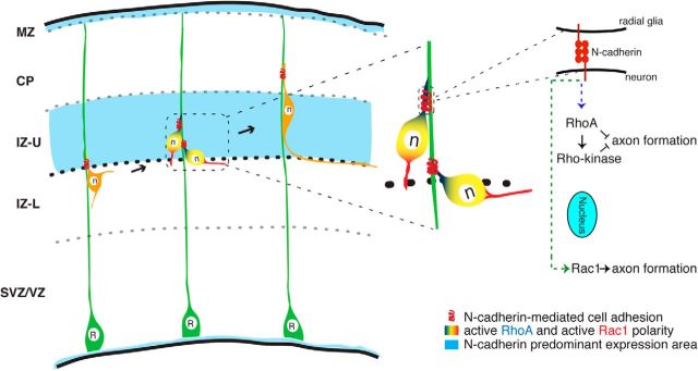
Figure 7.
Model depicting the mechanism by which pyramidal cells transit from multipolar to bipolar morphology and establish axon–dendrite polarity. The appearance of bipolar cell notably coincides with the appearance of the predominant expression of N-cadherin in the IZ. In the IZ, the N-cadherin-mediated radial glia–neuron interaction induces a polarized distribution of active RhoA at the contact site and active Rac1 in the opposing neurite. RhoA–Rho-kinase signaling in the contacting neurite inhibits axon formation; Rac1 in the opposite neurite promotes axon formation. R, Radial glial cell; n, neuron.

