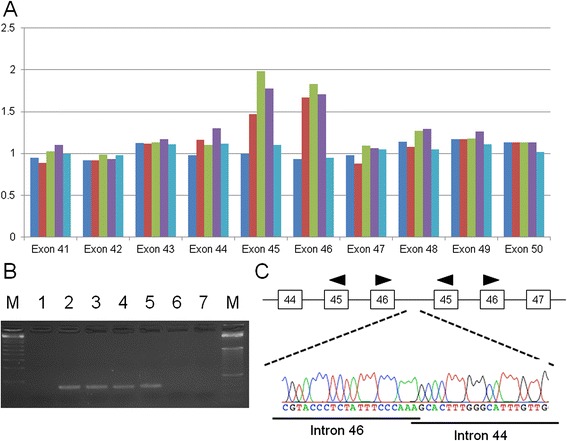
Fig. 4.

Exon duplication of the PKHD1 gene identified in the maternal allele. a Diagram of the MLPA results. Dark blue box, FHU14–046 (I-1, father); red box, FHU13–047 (I-2, mother); green box, FHU13–049 (II-1, healthy sibling); purple box, FHU13–068 (II-5, amniotic fluid); light blue box, normal healthy control. b Results of junction PCR analysis. Lane M, size markers; lane 1, FHU14–046 (I-1, father); lane 2, FHU14–047 (I-2, mother); lane 3, FHU13–049 (II-1, healthy sibling); lane 4, FHU13–051 (II-3, proband); lane 5, FHU13–068 (II-5, amniotic fluid); lane 6, normal healthy control; lane 7, water control. c Results of Sanger sequencing of the junction PCR product. The upper panel indicates the exon-intron structure around the junction
