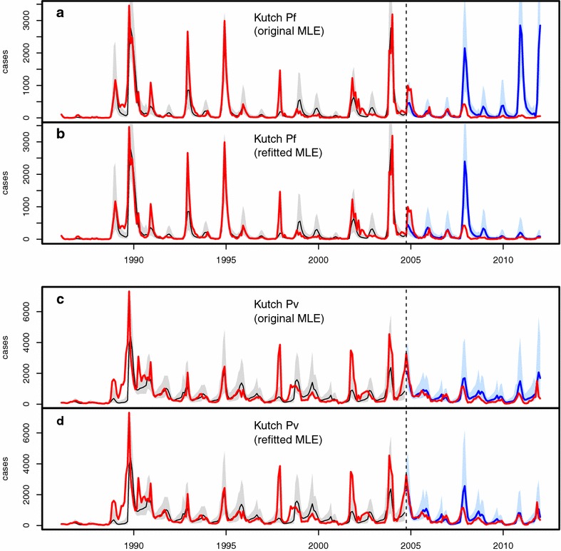
Fig. 3.

Comparison of model simulations with case data for Kutch. In all plots, monthly case data are shown in red, past simulations are in black (for the median of 10,000 simulations, with their corresponding 10–90 % confidence intervals, in light grey), and future simulations are in blue (10–90 % CI in light blue). a, b Present the results for falciparum malaria, and c, d for vivax malaria. Each panel consists of two plots, both including the same ‘past’ simulation but different future simulations corresponding respectively to those of method 1 (labelled ‘original MLE’) and method 2 (labelled ‘refitted MLE’)
