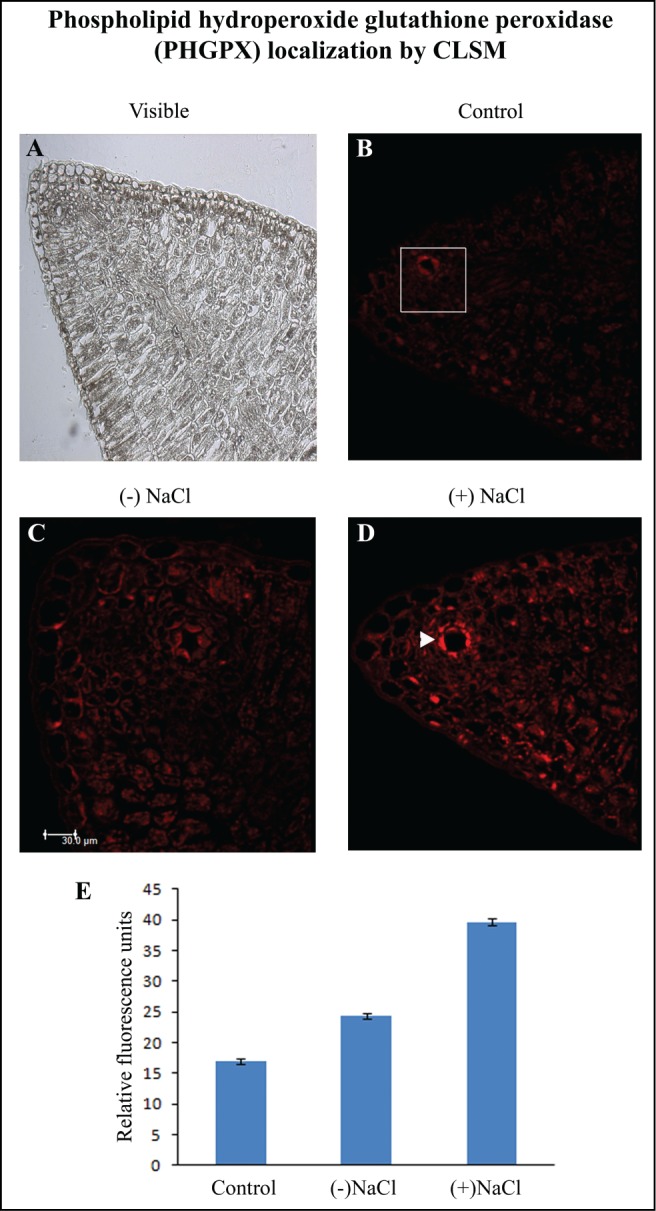
Figure 3.

Localization of phospholipid hydroperoxide glutathione peroxidase (PHGPX) by CLSM imaging using anti-GPX4 (PHGPX) antibody. (A) Visible image of 7 μm thick transverse sections of cotyledon from 2 d-old etiolated seedlings. Fluorescence micrographs of cotyledon transverse sections in the absence of anti-GPX4 (PHGPX) antibody (B), in the presence of anti-GPX4 (PHGPX) antibody in cotyledons from (−) NaCl seedling (C) and cotyledons from (+) NaCl seedlings (D); ▸ indicates secretory canal. (E) Quantification of fluorescence from the indicated zone. Magnification: 200×.
