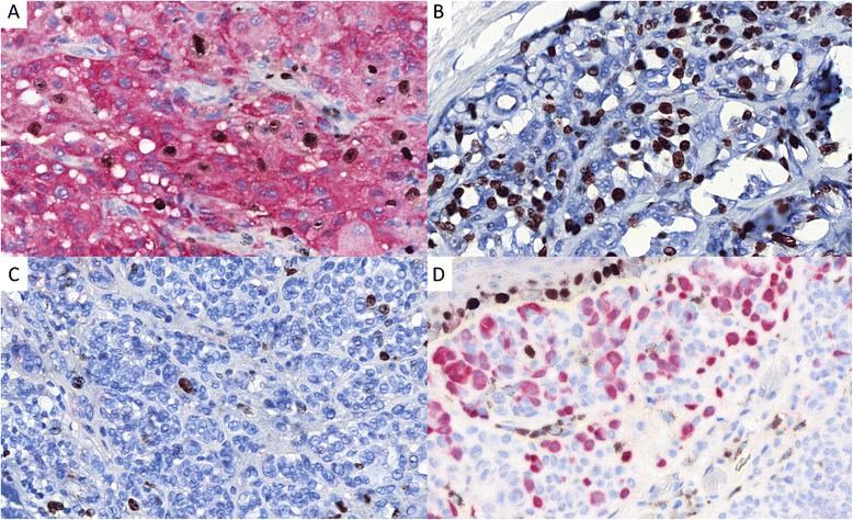
Fig. 3.

Illustrations of some Ki-67 (DAB) and p16 (Red) immunostaining results of the validation set of tumours (×400). a Diffuse p16 staining (score 0) within a melanoma with a Ki-67 index of about 15 % (score 3). b p16 negativity (score 3) within a melanoma with a Ki-67 index estimated at 50 % (score 4). c p16 negativity (score 3) within a melanoma with a Ki-67 index estimated at 3 % (score 1). D: About 40 % of nevocytes are stained with p16 (score 1) within a dermal nevus with a Ki-67 index of 1 % (note the Ki-67 stained basal keratinocytes as internal positive controls)
