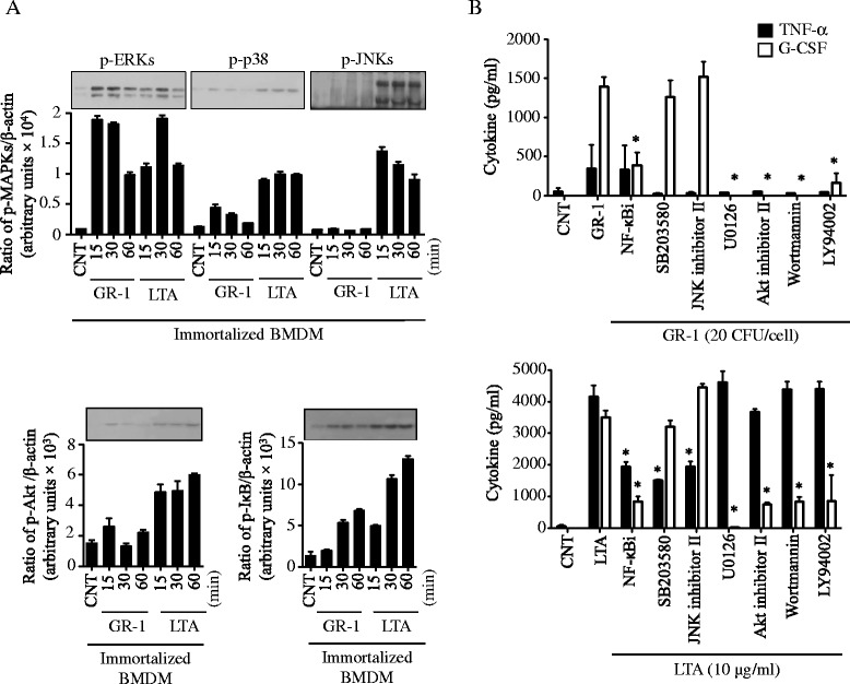
Fig. 3.

Activation of ERKs, NF-κB and Akt but not JNKs by GR-1 plays a key role in the preferential production of G-CSF over TNF-α. a Immortalized BMDMs were treated with live GR-1 (20 CFU/cell) or lipoteichoic acid (LTA; 10 μg/ml) for 15, 30 and 60 min. Phosphorylation of ERKs, p38, JNKs, Akt and IκB was analyzed by Western blots, using phospho-specific or β-actin antibodies. Bar graphs demonstrate band intensity quantification (ratio of protein phosphorylation to β-actin) using the NIH ImageJ program. Data shown as mean ± SEM [n ≥ 3]. b Immortalized BMDMs treated with GR-1 (20 CFU/cell) or lipoteichoic acid (LTA; 10 μg/ml) in the presence or absence of various inhibitors for NF-κB (NF-κBi; 10 μM), p38 (SB203580; 10 μM), JNKs (JNK inhibitor II; 250 nM), ERKs (U0126; 25 μM), Akt (Akt inhibitor II; 1 μM) and PI3K (wortmannin; 10 μM and PI3K-LY94002; 10 μM). Production of TNF-α (in 4 h) and G-CSF (in 24 h) was measured from cell culture media using ELISA. Data shown as mean ± SEM (n ≥ 3; *, p < 0.05 by Student’s t-test)
