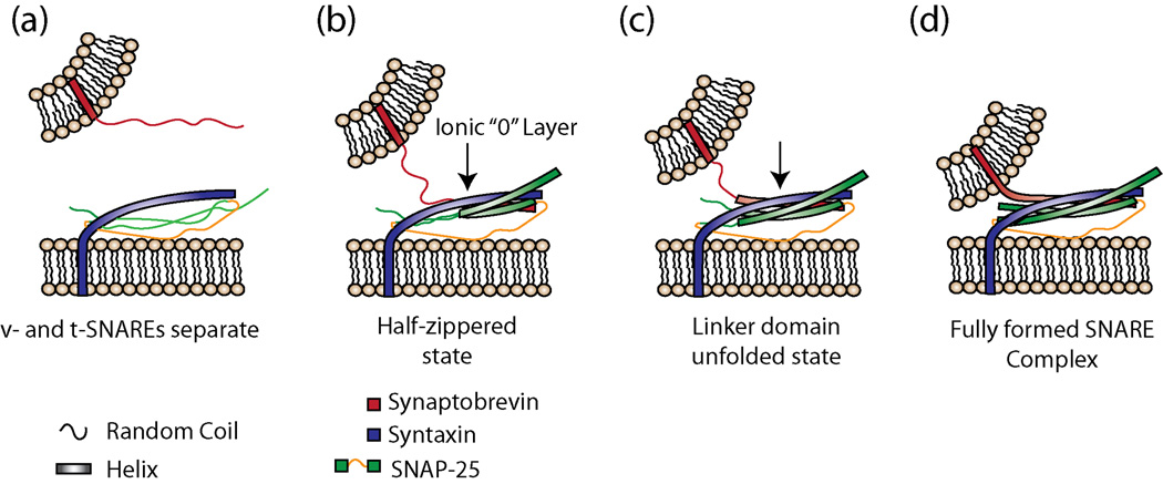
Fig 1.
SNARE complex structure formation during membrane fusion. (a) v- and t-SNAREs are separate. (b) Complex formation begins via association of the N-terminal regions of the individual SNARE proteins forming the half-zippered state [26,27**]. This association includes the ionic (0) layer. (c) A second intermediate state has been postulated [26], here called the partially unzipped state. (d) SNARE complex assembly and fusion complete. Here, the SNARE complex is drawn to reflect the conformation as observed in the crystal structure of the neuronal SNAREs [20]. It is still unclear at which point in these steps that membrane hemifusion and fusion occur.

