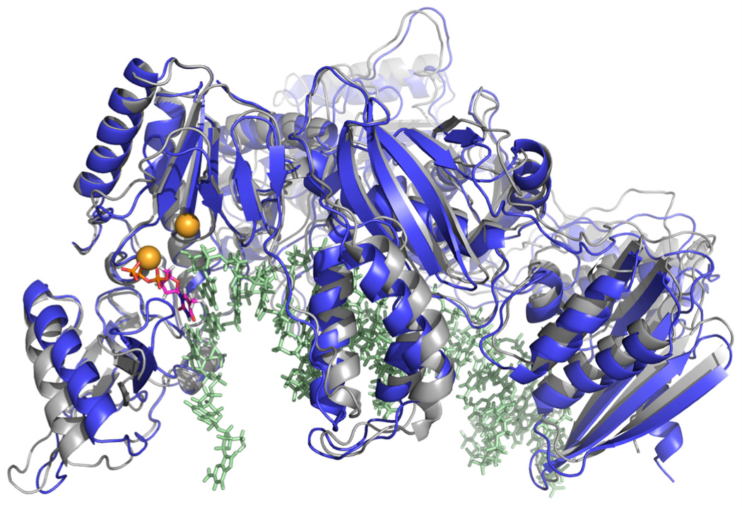
Figure 3.

Molecular structure of the HIV Reverse Transcriptase that is used in molecular dynamic simulations. The protein-DNA complex is shown in open (grey) and closed (blue) forms. Shown in colored spheres are Magnesium ions. The incoming nucleotide bound to the active site is shown with purple-orange sticks.
