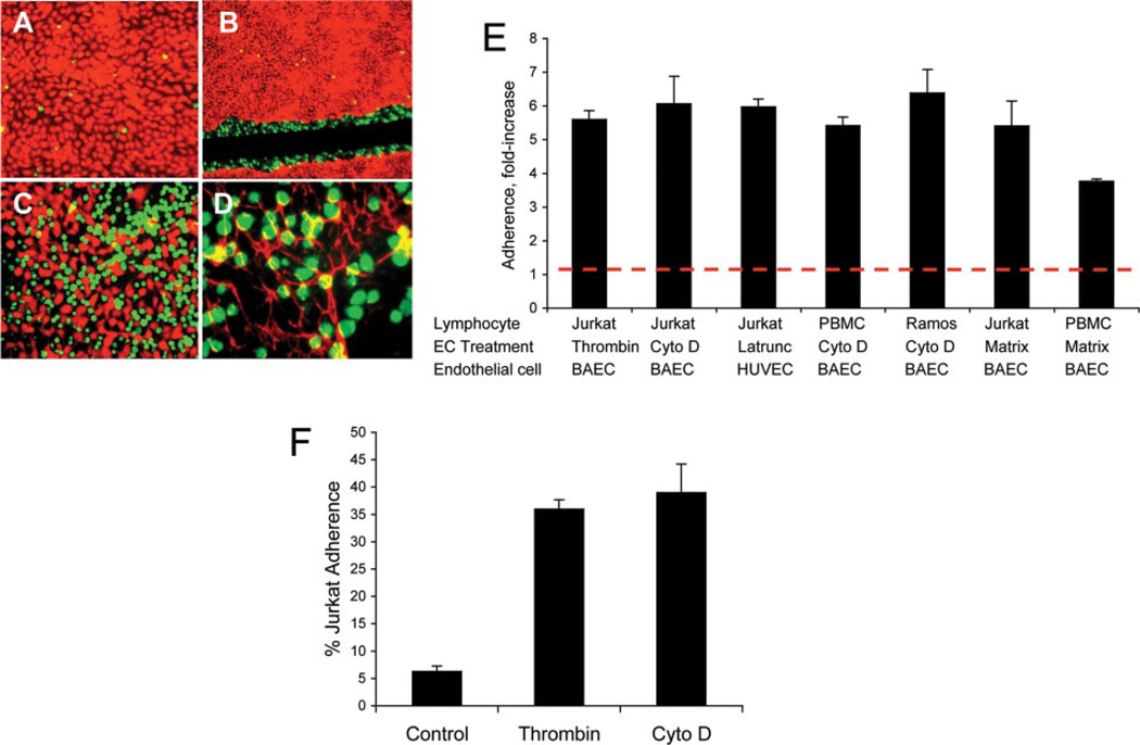
FIGURE 1.
Unstimulated leukocytes adhere to disrupted EC monolayers and the endothelial matrix. A, Adhesion of unstimulated Jurkat cells (green) to a confluent monolayer of BAEC (red) after 30 min. B, Adhesion of unstimulated Jurkat cells to the edges of a wounded BAEC monolayer. A confluent endothelial monolayer was wounded by scratching and the intervening matrix was removed by scraping and aspirating. C, Adhesion of unstimulated Jurkat cells to cytochalasin-retracted BAEC monolayer. D, Adhesion of unstimulated Jurkat cells to the BAEC-derived matrix. Fibronectin fibrils are visualized in red with an Ab to bovine fibronectin. Original magnification: ×10 (A and C); ×4 (B); and X20 (D). E, PBMC, Jurkat cells, or Ramos cells were allowed to adhere to the BAEC-, HUVEC-, or EC-derived matrix for 20 min at 37°C. EC treatments were: human thrombin, 10 U/ml for 10 min; cytochalasin D (Cyto D), 1 µM for 75 min; and latrunculin A, 1 µM for 50 min. The EC matrix was prepared by treating confluent EC monolayers with 20 mM NH4OH at 37°C for 5 min. x-Axis: fold- increase in adhesion compared with adhesion to the untreated endothelial monolayer of at least six independent experiments performed in triplicate wells. Dotted line represents adhesion to untreated endothelial monolayer. Values of p< 0.01 for all compared with controls. F, A representative experiment of Jurkat cell adhesion to thrombin- or cytochalasin D-retracted BAEC or untreated BAEC monolayer (control) is shown (n = 6 independent experiments). Data are the means of triplicate wells ± SD.

