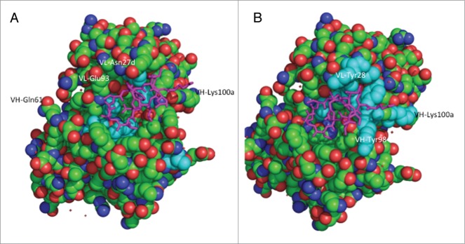
Figure 3.

Structure of hu5B3.v3 (space filling) in complex with epitope peptide (magenta tube).10 (A) Residues with greater than 5-fold loss in binding to HCV-gt1a-E2 when substituted with alanine colored cyan. (B) Residues included in affinity maturation library colored cyan.
