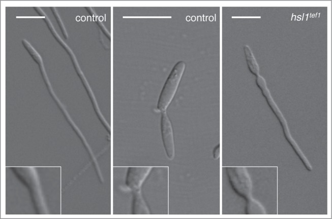
Figure 1.

Neck morphology in a wild-type (control) cell forming either an infective filament (left panel) or a bud (middle panel). Note the constriction observed in the bud neck (middle inset) in comparison with the absence of constrictions in the filament neck (left inset). In a strain that is not able to down-regulate the hsl1 expression during the formation of the infective filament (hsl1tef1) strikingly, the neck of the filament shows a constriction (right inset). Bar: 15 μm.
