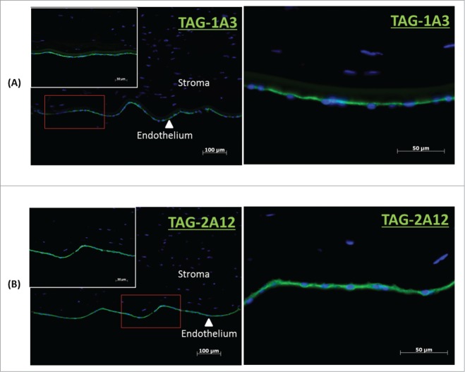
Figure 1.
Immunocytochemistry of frozen cornea tissue sections. (A) TAG-1A3 and (B) TAG 2A12. Primary antibodies were detected with Alexa Flour 488 secondary antibody and nuclei of cells were stained with DAPI. Both TAG-1A3 and TAG-2A12 had shown specific of staining for the endothelium layer in corneal tissue section.

