
Figure 5.
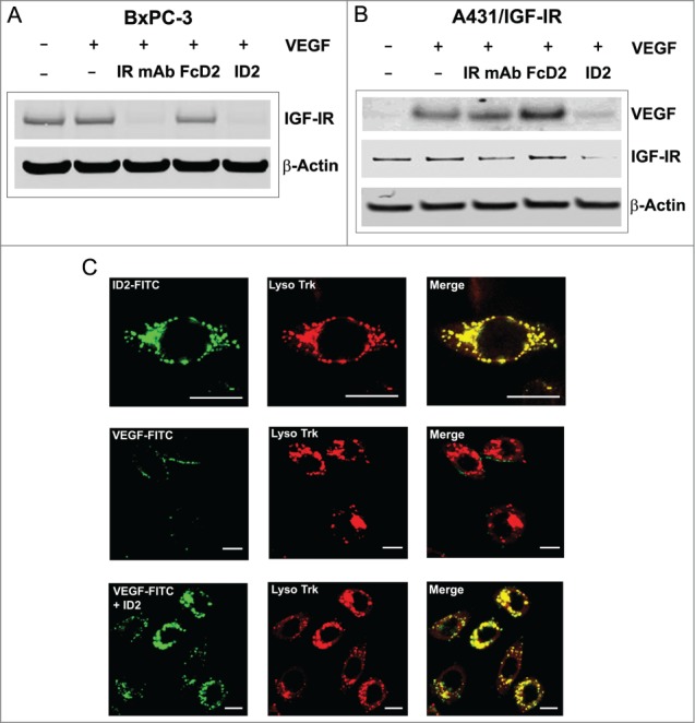
Evidence for internalization and degradation of both IGF-IR and VEGF by ID2 in vitro. (A) In BxPC-3 cells, ID2 at 20 nM induces IGF-IR internalization and degradation in the presence of 100 ng/mL VEGF by immunoblotting. IR mAb and FcD2 were used as controls. (B) In A431/IGF-IR cells, ID2 at 10 nM induces both surface IGF-IR and supernatant VEGF internalization/degradation in the presence of 400 ng/mL (10 nM calculated as a dimer) VEGF by immunoblotting. IR mAb and FcD2 were used as controls. (C) Fluorescence confocal microscopy analysis on the delivery of ID2 and VEGF to the lysosome in BxPC-3 cells: (top row) the co-localization of FITC-labeled ID2 with lyso tracker; (middle and bottom row) the co-localization of FITC labeled VEGF with the lysosome is dependent on ID2 treatment. Scale bar = 20 μm.