
Figure 1.
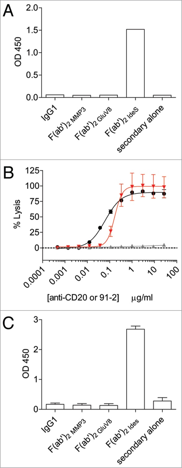
Generation of a monoclonal antibody specific for IdeS cleavage site in the lower hinge of human IgG1. (A) The fully rabbit anti-hinge mAb 91–2 bound to IdeS-generated F(ab’)2 fragments, but not to intact IgG1 and F(ab’)2 fragments generated with MMP-3 or GluV8 in a plate-based ELISA assay. Data are representative of two independent experiments (duplicate measurements per experiment). (B) CDC activity was measured using rituximab IgG1 (black circles), rituximab IgG1 F(ab’)2 IdeS (solid gray up triangles), and a fixed concentration of rituximab IgG1 F(ab’)2 IdeS (1 μg/ml) in the presence of serially-diluted 91–2 (solid red down triangles). Data are representative of two independent experiments (triplicate measurements per experiment). (C) The rabbit/human chimeric anti-hinge mAb 2095–2 displayed the same specificity as the fully rabbit mAb 91–2. Data are representative of two independent experiments (duplicate measurements per experiment).