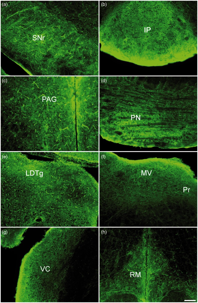
Figure 4.
AQP4 immunoreactivity within the brainstem, 10× images. (a) Substantia nigra pars reticulata (SNr). (b) Interpeduncular nucleus (IP). (c) Periaqueductal gray (PAG). (d) Pons (PN). (e) Lateral dorsal tegmental nucleus (LDTg). (f) Medial vestibular nucleus (MV) and nucleus prepositus (Pr). (g) Ventral cochlear nucleus (VC). (h) Raphe magnus (RM). Scale bar: 100 µm.

