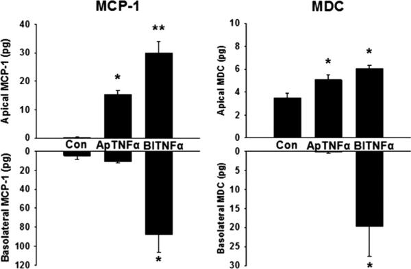
Fig. 4.

Intestinal epithelial cell chemokine production. Caco-2 cells were treated with apical or basolateral TNF-α or serum-free media. Monocyte chemoattractant protein 1 and MDC were measured in the apical and basolateral compartments by ELISA. *P < 0.05 vs. control, **P < 0.05 vs. control and apical treatment. n = 6.
