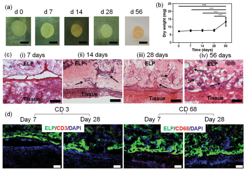
Figure 5.
Evaluation of degradation and biocompatibility of ELP hydrogels in vivo. ELP hydrogels were implanted into the dorsal subcutaneous space of rats. a) Macroscopic view on explanted ELP hydrogels 0, 7, 14, 28, and 56 d after implantation (scale bar = 5 mm). b) The in vivo degradation profile of ELP hydrogels (n = 5) over time based on dry weight measurements shows a significant gain in weight at day 56. c) Hematoxylin/eosin staining of subcutaneously implanted ELP hydrogels at postoperative days i) 7, ii) 14, iii) 28, and iv) 56 revealed progressive growth of host tissue into the implants, shown by the arrows (scale bar = 200 μm). d) Immunostaining of subcutaneously implanted ELP hydrogels at day 7 and 28 resulted in no local lymphocyte infiltration (CD3), and relevant macrophage detection (CD68) only at day 7, having disappeared by day 28 (scale bar = 100 μm). Green color in (d) represents the autofluorescent ELP, red color the immune cells, and blue color all cell nuclei (DAPI) (*** p < 0.001).

