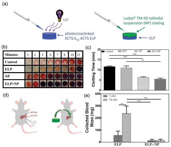
Figure 6.
Application of ELP hydrogels combined with silica nanoparticles as hemostats. a) A schematic of coating silica nanoparticle (NP; Ludox TM-50) solutions onto the photocrosslinked ELPs. b) A 96 well plate clotting time assay shows decreased clotting times when NP solutions (10 μL) are combined with the photocrosslinked ELPs (50 μL) (ELP + NP). c) Clotting times measured from the 96 well plate clotting time assay show a significant decrease in clotting time when NP was added. d) A schematic of NP-coated ELP hydrogel placement onto a lethal liver wound. e) In vivo blood loss after application of ELP versus ELP with NP coating showing a significant improvement when NP-coated ELPs are used to treat the wound (** p < 0.01, *** p < 0.001).

