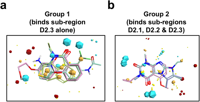
Figure 3. Consensus field maps for the two groups of small molecules that bound to p27-KID.
Blue and red field points indicate where electropositive and electronegative groups are favored, respectively, in the protein binding partner; gold field points indicate regions where hydrophobic interactions are favored; and yellow field points indicate regions where favorable van der Waals contacts are possible. The size of the field point increases with the magnitude of the favorable interaction energy. Two representative molecules from Group 1 (a) and Group 2 (b) are shown aligned to their respective consensus field maps.

