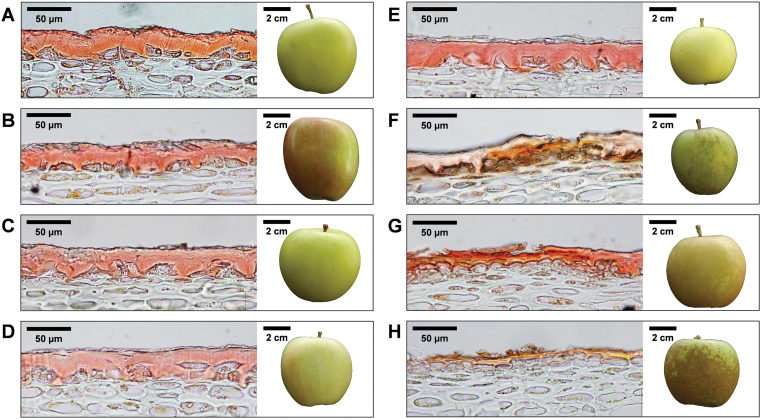
Fig. 4.
Histological analysis of apple skin tissues in a selected subset of the population. Light microscopy analysis of skin samples stained with Sudan IV are shown, together with photographs of whole apples. Sudan IV stains the cuticular lipids a pink/orange colour. (A, B) Parental lines ‘Golden Delicious’ (A) and ‘Braeburn’ (B), show regular cuticles. (C–E) Selected progeny with regular skin are shown: (C) G×B 3; (D) G×B 6; (E) G×B 11. (F–H) Selected progeny with compromised cuticles are shown: (F) G×B 30; (G) G×B 59; (H) G×B 90. Thinner, cracked cuticles are clearly visible in these lines, whereas whole apple photographs show the presence of russet on the surface of the fruit.

