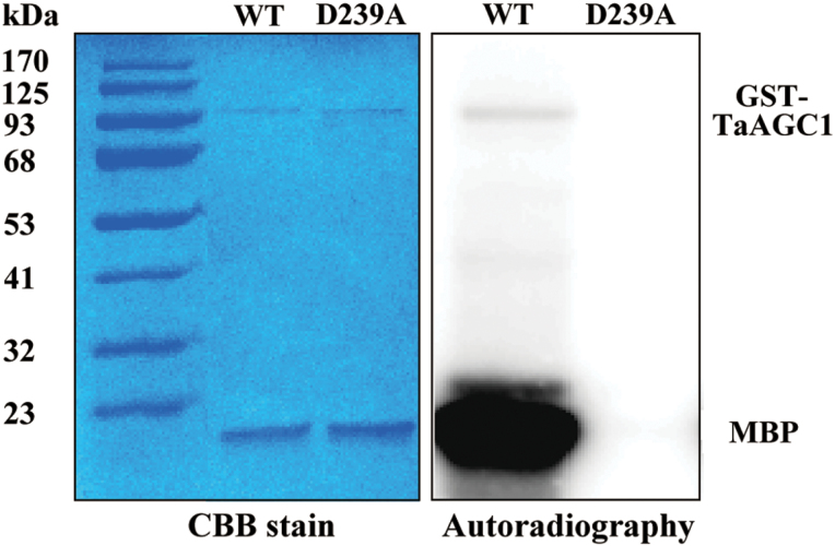
Fig. 3.
Analysis of the kinase activity of the TaAGC1 protein. The assays on autophosphorylation of TaAGC1 and myelin basic protein (MBP) phosphorylation by TaAGC1 were incubated for 30min in kinase buffer containing purified GST-TaAGC1 (WT) or GST-D239A mutant protein, the MBP substrate and [γ-32P] ATP. The phosphorylated proteins were separated by SDS-PAGE and visualized by autoradiography. After autoradiography, the right proteins were confirmed by Coomassie Brilliant Blue (CBB) staining. (This figure is available in colour at JXB online.)

