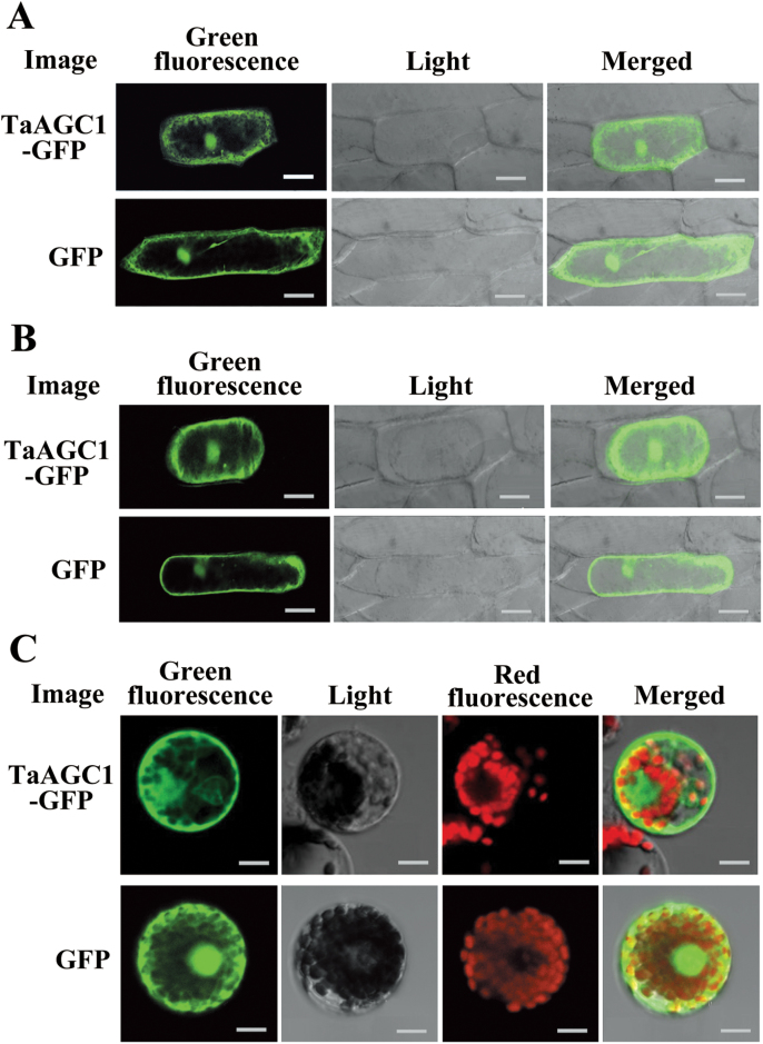
Fig. 4.
Subcellular localization of the wheat (Triticum aestivum) AGC kinase TaAGC1-green fluorescent protein (GFP) fusion protein in onion and wheat. (A) The fused TaAGC1-GFP (upper panel) and control GFP (lower panel) in onion epidermal cells. (B) The fused TaAGC1-GFP (upper panel) and control GFP (lower panel) in plasmolysed onion epidermal cells. Bars, 50 μm (A, B). (C) The fused TaAGC1-GFP (upper panel) and control GFP (lower panel) in wheat protoplasts. Bar, 10 μm. (This figure is available in colour at JXB online.)

