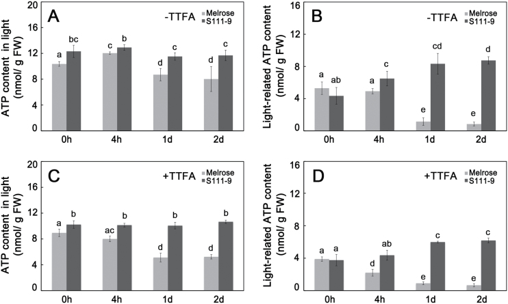
Fig. 4.
ATP contents of leaves under 150mM salt stress. The leaves used for these measurements were the same as those used for the Chl fluorescence and dark re-reduction of P700+ analysis shown in Fig. 3. All data were determined in Melrose and S111-9 under salt stress after 0h, 4h, 1 d, and 2 d. (A) ATP content in light in the absence of 100 μM TTFA (an nhibitor that binds ferredoxin); (B) light-related ATP content in the absence of 100 μM TTFA, as calculated by subtracting the ATP content in darkness from the ATP content in light. The samples for ATP content in darkness were collected and kept in darkness for 24h, and reflect the ATP produced by oxidative phosphorylation. The light-related ATP content reflects the ATP accumulation produced by photophosphorylation. (C) ATP content in light in the presence of 100 μM TTFA; (D) light-related ATP content in the presence of 100 μM TTFA. Bars represent the mean ±SD of four independent experiments. Different letters indicate significantly different values (P<0.05) by Tukey’s test.

