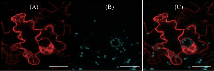
Fig. 5.
Localization of OsCOMT. (A) Red fluorescence of OsCOMT-mCherry and (B) chlorophyll (Chl) autofluorescence. (C) The two fluorescence images were merged (A+B). Tobacco (N. benthamiana) leaves were infiltrated with Agrobacterium harbouring the XVE-inducible OsCOMT-mCherry binary vector, as described in Materials and methods. Bars, 40 μm. (This figure is available in colour at JXB online.)

