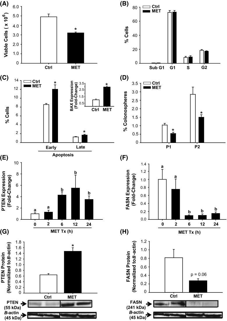
Fig. 2.
Metformin (MET) altered growth and survival parameters and modified PTEN and FASN expression of HCT116 cells. a HCT116 cell viability after treatment with 60 μM MET for 48 h. b Treatment with MET for 48 h had no effect on cell cycle distribution, as determined by propidium iodide (PI) staining and subsequent FACS. c MET increased the number of apoptotic cells relative to control. Proportion of apoptotic cells was determined by Annexin V staining and FACS (“Materials and methods”), and the induction in apoptosis was associated with an increase in expression of the pro-apoptotic BAX gene (inset). d MET lowered P1 and P2 colonosphere formation of HCT116 cells. Results (a–d) are mean ± SEM; *P < 0.05 relative to control, with n = 2–3 independent experiments and each experiment performed in quadruplicates. e, f Transcript levels of PTEN and FASN genes at specific time points were quantified (QPCR) in control and MET-treated cells. Data are mean ± SEM of fold-change in normalized expression (n = 5 samples/group), relative to control. g, h Representative Western blots of whole cell extracts after treatment with MET. PTEN and FASN protein levels were compared to control (vehicle-treated cells). Each lane represents an individual treatment sample and contained 50 μg of total protein. Immunoreactive bands were quantified by densitometry; expression values (mean ± SEM) were normalized to those of the loading control β-actin and are presented as histograms. *P < 0.05 (relative to control)

