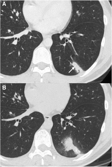
Fig. 1.

Computed tomography scan of the chest. a Chest computed tomography showed an irregularly shaped solitary nodule in the periphery of the left lower lobe, with microcalcifications (arrow) and pleural indentation. b After 2 weeks, the nodules became enlarged, around which ground glass opacity appeared
