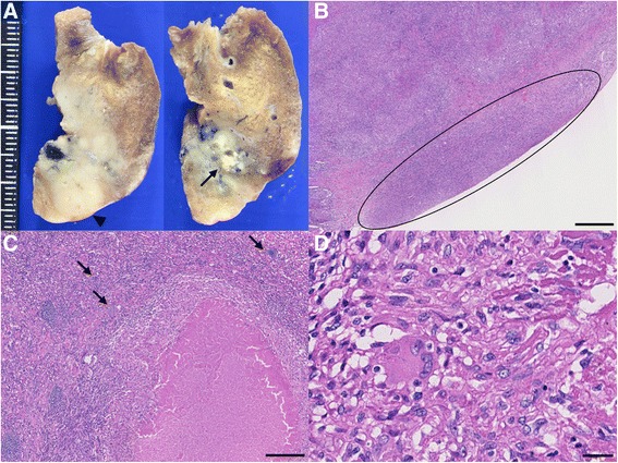
Fig. 3.

Photograph and photomicrographs of the lung. a Photograph of a cross-sectional specimen in the resected lung showing a granuloma with caseating necrosis (arrow) and pleurisy (arrowhead). b Photomicrograph showing pleurisy over the visceral pleurae (circle; bar 500 μm), which revealed granulomatous infiltration. c-d Photomicrograph showing an epithelioid granuloma with necrosis and calcification (arrows) (C; bar 250 μm, D; bar 25 μm)
