
Fig. 2. Porphyromonas gingivalis induces nuclear accumulation of FOXO1.
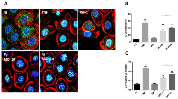
A. Fluorescent confocal microscopy of TIGK cells infected with P. gingivalis 33277 (Pg) at MOI 10 or 100 for 2 h. Control cells were non-infected (NI). Cells were fixed and probed with FOXO1 antibodies (green). Actin (red) was stained with Texas Red phalloidin, and nuclei (blue) stained with 4′,6-diamidino-2-phenylindole (DAPI). Tumour necrosis factor (TNF, 5 ng ml−1) was a positive control for FOXO activation and insulin-like growth factor (IGF-1, 50 ng ml−1) was a negative control. Cells were imaged at magnification ×63 and shown are representative merged images of projections of z-stacks (10 slices/z stack) obtained with Volocity software.
B and C. Nuclear localization of FOXO1 calculated by colocalization of FOXO1 staining with nuclei (B), and Pearson’s correlation coefficient (C) from images in A (n = 200 cells) using Volocity software. Results are means ± standard deviation; *P < 0.05; **P < 0.01.