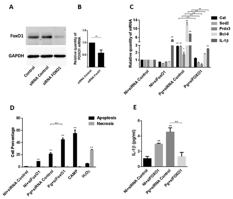
Fig. 5. FOXO1 knockdown suppresses TIGK responses to P. gingivalis.
A. TIGK cells were transfected with siRNA to FOXO1 or scrambled siRNA (siRNA control). Control cells were non-transfected. Cell lysates were immunoblotted with FOXO1 antibodies or GAPDH antibodies as a loading control.
B. FOXO1 mRNA levels in transfected TIGK cells (as in A) were measured by qRT-PCR. Data were normalized to GAPDH mRNA and are expressed relative to the siRNA control. Results are means ± standard deviation (SD); n = 3; **P < 0.01.
C. Transfected TIGK cells were infected with P. gingivalis 33277 (Pg) MOI 10 for 2 h and Cat, Sod2 Prdx3, Bcl-6 and IL-1β mRNA levels were measured by qRT-PCR. Data were normalized to GAPDH mRNA and are expressed relative to non-infected (NI) siRNA controls. Results are mean ± SD; n = 3; *P < 0.05 *; **P < 0.01.
D. Transfected TIGK cells were infected with P. gingivalis 33277 (Pg) MOI 10 for 2 h or left uninfected (NI) and the level of apoptosis and necrosis determined by staining with AnnexinV and SytoxGreen, respectively, followed by flow cytometry. Campthothecin (CAMP, 10 μM) or H2O2 (0.3%) were positive controls for apoptosis or necrosis respectively. Results are mean ± SD; n = 3; **P < 0.01.
E. Transfected TIGK cells were infected with P. gingivalis 33277 (Pg) MOI 10 for 2 h, or left uninfected (NI), and IL-1β levels in culture supernatants determined by ELISA. Results are mean ± SD; n = 3; **P < 0.01.

