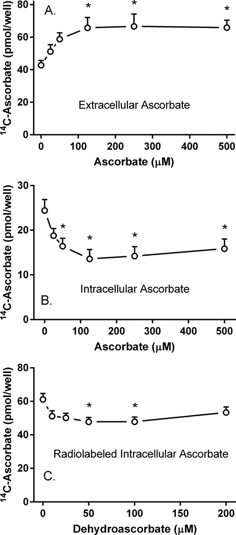
Figure 3. Effects of intra- and extracellular unlabeled ascorbate on radiolabeled ascorbate efflux.
Pericytes in 12-well plates were incubated for 60 min with 10 µM radiolabeled ascorbate, the medium was aspirated, and the cells were rinsed twice in KRH containing 5 mM D-glucose. The cells were incubated with 1 ml of glucose-KRH containing the indicated concentration of unlabeled ascorbate. After 60 min, radiolabeled ascorbate was measured in the medium (Panel A) or in the cells (Panel B) as described in Materials and methods. Results are shown from 6 experiments, with an “*” indicating p < 0.05 compared to the sample not treated with unlabeled ascorbate. Panel C. Pericytes were loaded with 10 µM radiolabeled ascorbate for 20 min in complete medium, followed by addition of the indicated concentration of DHA. After another 30 min at 37°C, the medium was removed and the cells were rinsed twice in 37°C glucose-KRH. Following addition of 1 ml of glucose-KRH at 37°C, efflux was measured at 30 min as described in Materials and methods. Results are shown from 6 experiments, with an “*” indicating p < 0.05 compared to the sample not treated with DHA.

