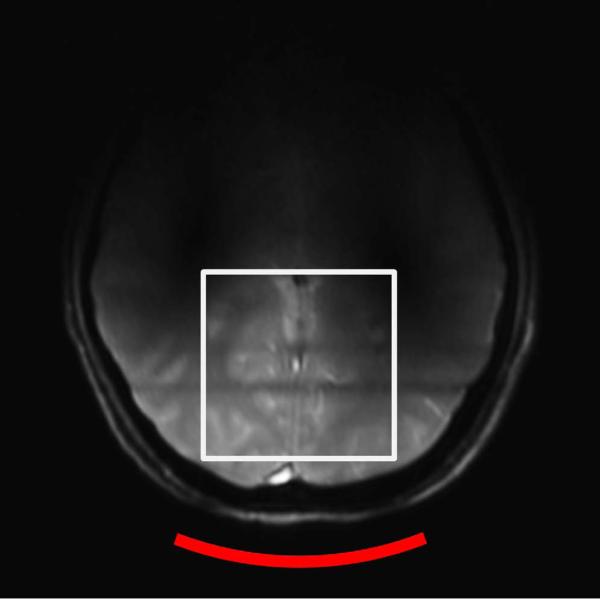
Figure 2.
An axial image of the human brain obtained on the 7 T scanner using a gradient echo based three-plane localizer sequence and the quadrature surface coil. TR = 8.6 s, TE = 2.9 s, FOV = 28 cm, and no. of average = 1. The white square box illustrates the location of the cubical shim region (6 × 6 × 6 cm3).

