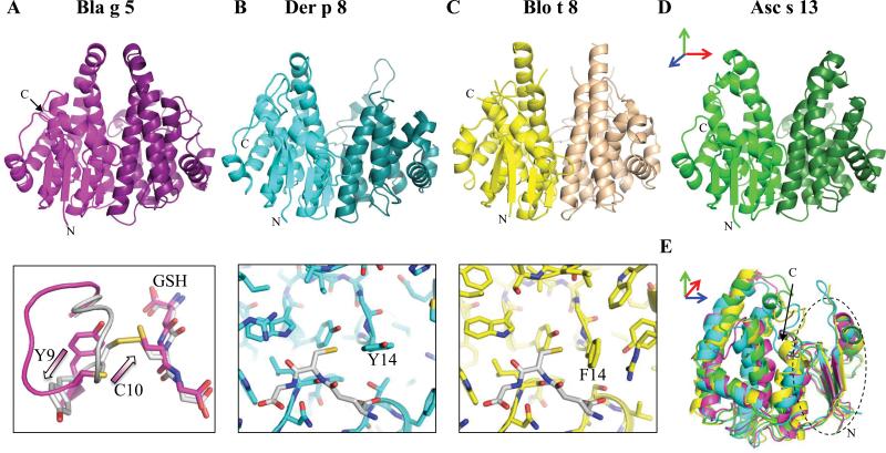
Figure 1. GST Allergen Structures.
A-C (top) and D) Ribbon diagrams of the dimer. E) Superposition of monomers, and N-terminal thioredoxin-like sub-domain (dashed oval). Two active site conformations of Bla g 5, and the active site loop of Der p 8 and Blo t 8 (GSH molecule in white) are shown in bottom of A, B and C, respectively. Arrows in three colors indicate how the structures in A-D are rotated with respect to E. Black arrows in A and E show the C-termini.

