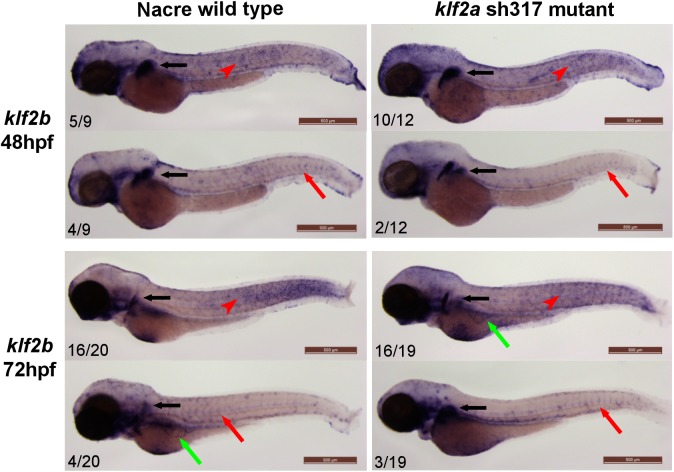
Fig 8. klf2b expression patterns do not differ between wildtype and klf2a sh317 mutants.
klf2b mRNA is detectable in the developing pectoral fin bud (black arrows). Most of the embryos examined exhibit klf2b mRNA presence on the surface of the embryos representing epidermal cells as described before [17, 48] (red arrowheads). A small proportion of both wildtype and klf2a sh317 mutant embryos show vascular staining in the ISVs (red arrows) and/or in the subintestinal veins (green arrows). There were no differences in staining patterns and especially in the level of vascular staining observed between wildtype and klf2a sh317 mutant embryos. Figures in bottom left corner of each image indicate the number of embryos with similar staining patterns out of total number of embryos examined. Scale bar = 500μm.

