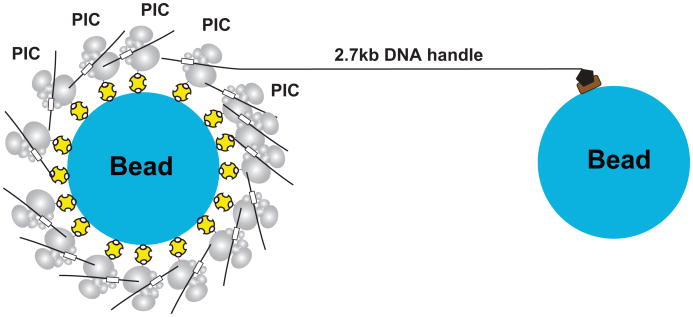
Extended Figure 2. Schematic diagram showing assembly of dumbbells.
PICs were attached to one bead via biotin-avidin linkages (yellow). To form dumbbell tethers, the other end of a small fraction of the PICs (4%) had digoxigenin linkages that could be tethered to anti-digoxigenin-coated beads (black and brown) via a 2.7 kb DNA handle. PICs not involved in tether formation served to increase the local concentration of PIC components.

