Figure 4. Tse6 Participates in a Multi-protein Complex and Binds Helix D of EF-Tu through Residues N-Terminal to Its Toxin Domain.
(A) Tse6 is a membrane protein that is solubilized by VgrG1. Western blot analysis of the soluble (S) and membrane (M) fractions of the indicated P. aeruginosa strains. Tse1 and OprF serve as soluble and membrane controls, respectively.
(B) Silver-stained SDS-PAGE analysis of proteins enriched by anti-VSV-G immunoprecipitation from P. aeruginosa strains encoding Tse6 (parental) and Tse6-V. The labels indicate the identities of proteins that specifically co-precipitate with Tse6-V as determined by MS. In addition to their monomeric forms, VgrG1 and Tse6 form a high-molecular-weight complex that is resistant to heat and SDS denaturation.
(C) A 17-amino-acid segment of Tse6 mediates interaction with EF-Tu. Coomassie-stained SDS-PAGE analysis of purified Tse6 truncations. All truncations were expressed with Tsi6 and assessed for co-purification with endogenous EF-TuEC.
(D) Overall structure of the Tse6265-CT–EF-TuPA complex. Secondary structure elements involved in the interaction are labeled.
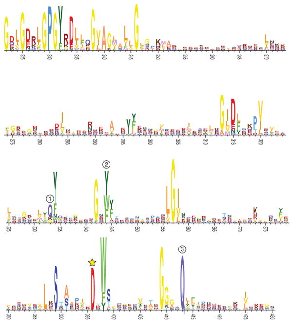
(E) The Tse6 activation loop harbors Asp396 and rotates toward the active site of Tse6 in the Tse6265-CT –EF-TuPA structure relative to its position in the Tse6282-CT–Tsi6 structure. Dots denote a disordered segment (amino acids 400–408) of Tse6282-CT that was not modeled.
(F) Tse6282-CT D396A exhibits significantly reduced NAD(P)+ glycohydrolase activity. Rate of NAD+ (left) and NADP+ (right) consumption by purified Tse6282-CT D396A.
(G) Asp396 is critical for Tse6-based intercellular toxicity. Growth competition experiments between the indicated P. aeruginosa donor and recipient strains. Donor and recipient strains were mixed 1:1, grown for 24 hr on solid media, and differentiated using blue/white screening.
(H) Helix D of EF-Tu is the site of interaction for both Tse6265-CT (left) and the guanine exchange factor EF-Ts (right). In all panels, error bars represent ± SD (n = 3).
See also Figures S3 and S4 and Tables S1–S3.

